Figure 3.
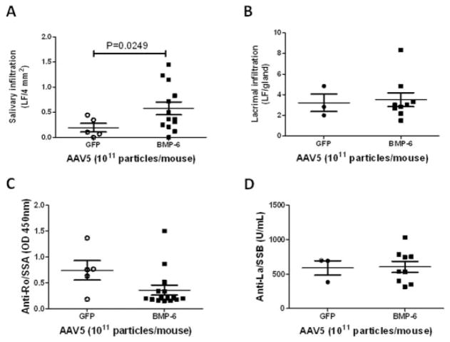
Lymphocytic infiltration and autoantibody production in AAV5-Bmp6– and AAV5-GFP–treated mice. Lymphocytic infiltrates were determined as described in Materials and Methods, and a focus score of lymphocytic foci (LF) per 4 mm2 was assigned. A, The focus score in salivary glands from AAV5-Bmp6–treated mice (n = 13) was significantly increased compared with that in salivary glands from AAV5-GFP–treated mice (n = 5). B, After delivery of vector to the salivary glands, the lacrimal gland focus score did not differ significantly (P = 0.8215) between the AAV5-Bmp6–treated group (n = 9) and the AAV5-GFP–treated group (n = 3). C and D, Serum levels of anti-Ro/SSA (C) and anti-La/SSB (D), as determined by enzyme-linked immunosorbent assay, did not differ significantly (P = 0.0685 for anti-Ro/SSA, P = 0.0987 for anti-La/SSB) between groups (n = 5 in the AAV5-GFP–treated group, n = 15 in the AAV5-Bmp6–treated group in C; n = 3 in the AAV5-GFP–treated group, n = 9 in the AAV5-Bmp6–treated group in D). Bars show the mean ± SEM. P values were determined by Student’s unpaired t-test. See Figure 2 for other definitions.

