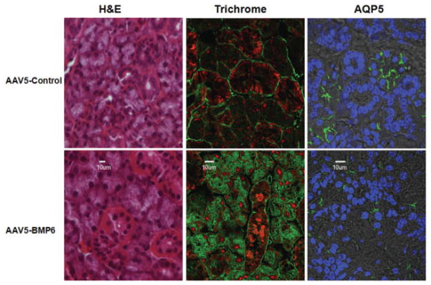
Figure 4.
Morphologic changes in the salivary glands of AAV5-Bmp6–treated mice. Changes in morphology and in protein expression or distribution were assessed by hematoxylin and eosin (H&E) or trichrome staining or immunofluorescence confocal imaging for aquaporin 5 (AQP-5). Each image is representative of 4 samples tested. No gross changes in morphology were observed with H&E staining. Trichrome staining revealed a redistribution of the extracellular matrix material in AAV5-Bmp6–treated mice compared with AAV5-GFP–treated controls. AQP-5 appeared to be less well defined on the apical surfaces in the AAV5-Bmp6–treated mice compared with the AAV5-GFP–treated mice. Original magnification × 40 (H&E) or × 100 (trichrome and AQP-5). See Figure 2 for other definitions.

