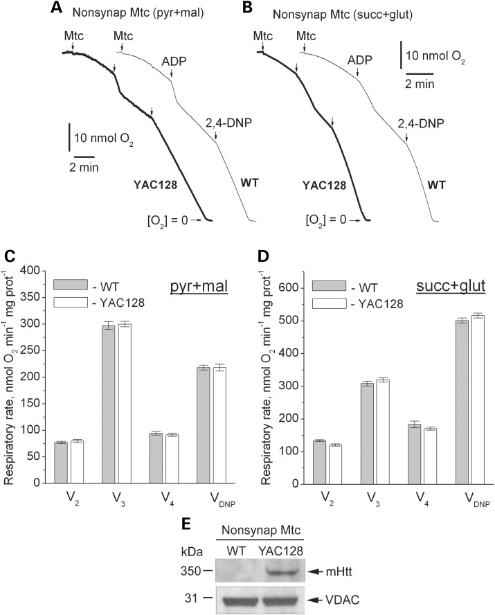
Figure 4.
Respiratory activity of brain non-synaptic mitochondria isolated from 2-month-old WT (thin traces) and YAC128 (thick traces) mice. In (A and B) representative traces for mitochondrial O2 consumption. Where indicated, non-synaptic mitochondria (Mtc), 200 μm ADP and 60 μm 2,4-dinitophenol (2,4-DNP) were added. In (A), incubation medium was supplemented with 3 mm pyruvate (pyr) and 1 mm malate (mal). In (B), incubation medium was with 3 mm succinate (succ) and 3 mm glutamate (glut). In (C and D) statistical analysis of respiratory rates. Data are mean ± SEM, N = 10. In (E) western blot, indicating the presence of mHtt in non-synaptic mitochondria from YAC128 mice. VDAC was used as a loading control.

