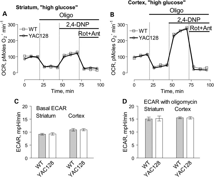
Figure 9.
OCR and ECAR of cultured neurons from YAC128 and WT mice: ‘high glucose conditions’. In these experiments, we used striatal and cortical neurons derived from postnatal Day 1 YAC128 and WT mice. The cells were grown for 9 days in vitro (9 DIV) before measurements. The bath solution contained 10 mm glucose and 15 mm pyruvate to accentuate mitochondrial respiration (22). Where indicated, cells were treated with 1 µm oligomycin (Oligo), 60 µm 2,4-dinitrophenol (2,4-DNP), 1 µm rotenone (Rot) and 1 µm antimycin A (Ant). In (A and B), OCR of striatal and cortical neurons, respectively. In (C and D), ECAR of striatal and cortical neurons. The OCR and ECAR were measured with Seahorse XF24 flux analyzer (Seahorse Bioscience, Billerica, MA, USA) at 37°C with 105 cells per well. Data are mean ± SEM, N = 7.

