Figure 4.
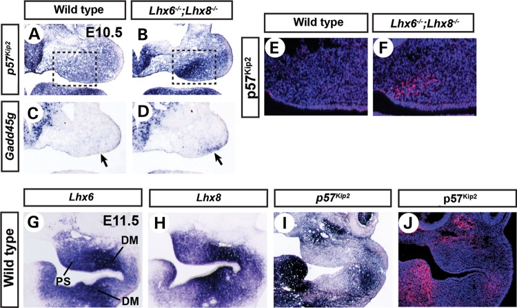
Repression of p57Kip2 by Lhx6 and Lhx8 in the palate area of the maxillary arches. (A–D, G–I) Coronal sections of the head of E10.5 (A–D) or E11.5 (G–I) embryos processed by RNA in situ hybridization. The right half of the face is shown. The boxes in A and B highlight the strong up-regulation of p57Kip2 in the prospective palate area. The arrows in C and D point to the up-regulation of Gadd45g in the oral-lateral domain of Lhx6−/−;Lhx8−/− mutant maxillary arches. (E, F and J) Coronal sections of the head of E10.5 (E and F) or E11.5 (J) embryos stained for nuclei (blue) and p57Kip2 protein (red). E and F are equivalent to the boxed areas in A and B. DM, dental mesenchyme.

