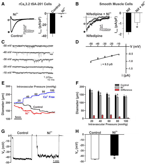
Figure 1. Effects of Ni2+ on CaV3.2 currents, myogenic tone, and membrane potential (VM).
A, Representative traces and summary data of inward currents in CaV3.2-transfected tSA-201 cells in the absence and presence of Ni2+ (CaV3.2 blocker, 50 μmol/L). A voltage step from −90 to −10 mV was used to evoke inward Ba2+ current (n=5; *P<0.05, paired t test). B, T-type current in rat cerebral arterial smooth muscle before and after Ni2+ (50 μmol/L). Experiments were performed in the presence of nifedipine (200 nmol/L) to block L-type Ca2+ channels. A voltage step from −90 to 0 mV was used to elicit inward current (n=8; *P<0.05, paired t test). C and D, Single-channel recordings and summary current–voltage relationship (n=5; slope conductance=8.5 pS) of T-type Ca2+ currents in cerebral arterial myocytes. On-cell recording was performed at −50 to −10 mV in the presence of nifedipine (200 nmol/L), 60 mmol/L Ca2+, and 50 mmol/L TEA-Cl (tetraethylammonium chloride) to block K+ channels. E and F, Rat middle or posterior cerebral arteries were pressurized from 20 to 100 mm Hg, whereas diameter was monitored under control conditions, in the presence of Ni2+ (50 μmol/L) and in Ca2+-free medium. Representative traces (E) and summary data (F) display augmented arterial tone in response to Ni2+ (n=7; *P<0.05, paired t test). G and H, Arterial VM in pressurized cerebral arteries (60 mm Hg) in the absence and presence of Ni2+ (50 μmol/L). Illustrative traces (G) and summary data (H) reveal the depolarizing effect of Ni2+ (n=6; *P<0.05, paired t test).

