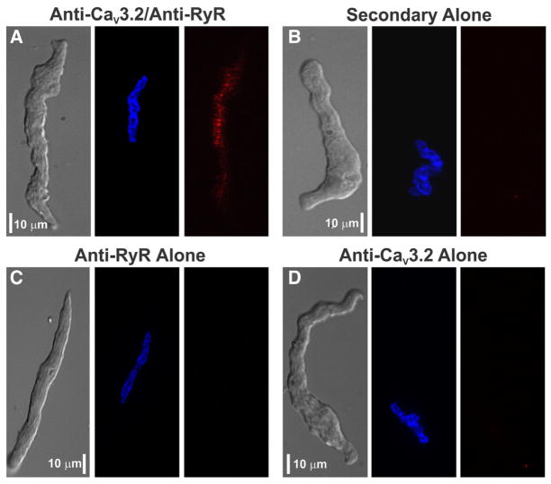
Figure 4. Proximity ligation assay of CaV3.2 and ryanodine receptor 2 (RyR2) in rat cerebral arterial smooth muscle cells.
A, A gallery representation reveals the presence of red fluorescent product consistent with CaV3.2 and RyR colocalization within 40 nm of one another. Nuclei were labeled with Hoechst 33342 (blue). B, Assay was performed with no primary antibodies. C and D, Assay controls were developed with 1 primary antibody. Scale bars are 10 μm, and optical section depth in each image is 0.3 to 0.5 μm. Photomicrographs are representative of ≈10 to 20 smooth muscle cells, and the assay was tested 2 to 3 times for each panel.

