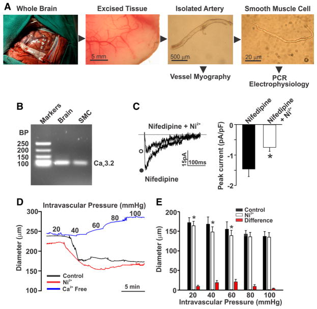
Figure 8. Expression and function of CaV3.2 in human cerebral arteries.
A, Brain tissues were excised from patients undergoing lobectomy. Whole cerebral arteries and cerebral arterial smooth muscle cells (SMCs) were subsequently isolated for experimental assessments. B, Polymerase chain reaction (PCR) analysis of whole brain and isolated SMCs highlights the presence of CaV3.2. Data are representative of cells obtained from 2 human subjects. C, A voltage step from −90 to 0 mV was used to monitor inward Ba2+ current (n=6 cells from 4 subjects) in human cerebral arterial SMCs in the absence or presence of Ni2+ (50 μmol/L; *P<0.05, paired t test). Currents were monitored in the presence of nifedipine (200 nmol/L) to block L-type Ca2+ channels. D and E, Human cerebral arteries were pressurized from 20 to 100 mm Hg while diameter was sequentially monitored under control conditions and in the presence of Ni2+ (50 μmol/L) or in Ca2+-free media (n=4 arteries from 3 subjects; *P<0.05, paired t test). BP indicates base pairs.

