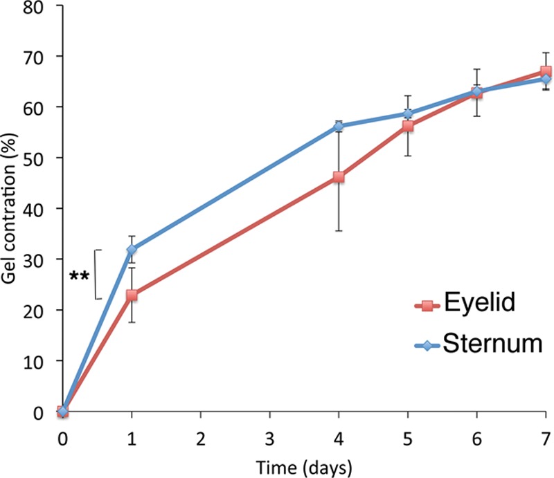
Fig. 2.

Presternal cutaneous fibroblasts display increased early contraction of a collagen matrix after serum stimulation when compared with matching eyelid cells. Fibroblasts from the eyelid (red), or from the presternal area (blue), were embedded in free-floating collagen matrices and monitored daily for 7 days. Contraction is expressed as the percentage decrease in the gel area, relative to the area at time 0. Shown is the mean result of at least 3 experiments run in triplicate; error bars indicate standard error. ** indicates significant difference, P < 0.01.
