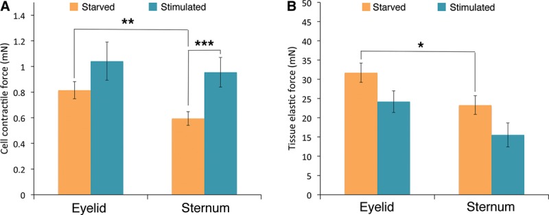
Fig. 3.

Presternal and eyelid cutaneous fibroblasts differ in their cell-force profiles: eyelid fibroblasts and presternal cells were cultured in collagen gels to generate 3D tissue-like matrices, which were then starved overnight in serum-free medium. Cellular force generation and tissue matrix rigidity were determined before (starved) and after stimulation with 20% FBS for 15 minutes (stimulated). A, Eyelid cutaneous fibroblasts display greater intrinsic baseline cellular contractile force when compared with matching presternal fibroblasts under resting conditions (**, P < 0.01), but only presternal cells showed a significant increase in contractile force following stimulation (***, P < 0.001). B, Matrices prepared with eyelid cutaneous fibroblasts displayed greater elastic force than those made with presternal cells (*, P < 0.02), with no change after serum stimulation.
