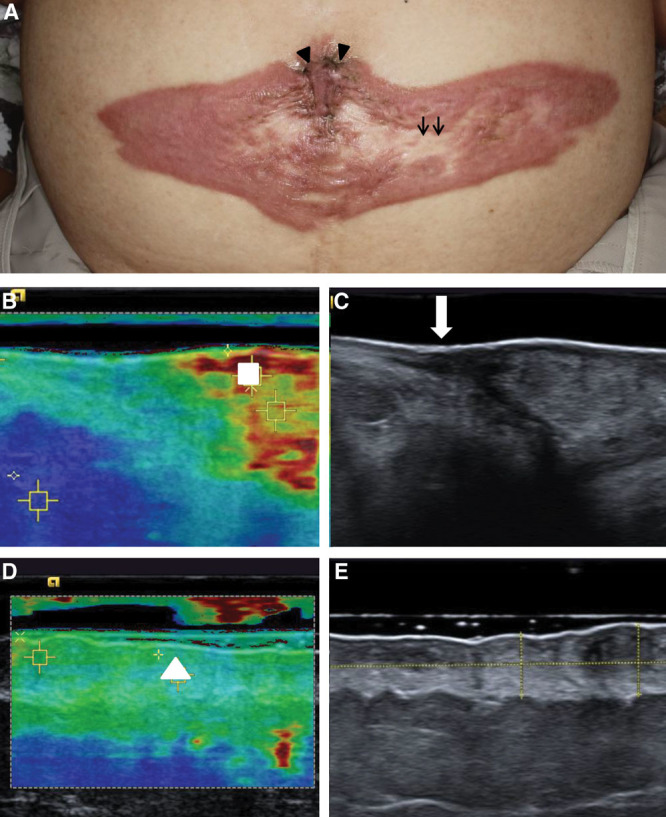
Fig. 1.

Case 1. Abdominal keloid in a 76-year-old woman (A). The keloid contained a hypertrophic area (arrowhead) and mature area (arrow). Ultrasound elastography and B-mode image of the hypertrophic area (B). The white arrow shows the boundary between the normal skin and the keloid. The velocity on elastography measured in the white square in the hypertrophic area was v = 7.09 m/s. Image of the mature lesion (C). The velocity on elastography measured in the white triangle was v = 3.12 m/s.
