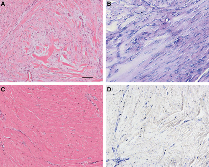
Fig. 2.

HE staining of the hypertrophic area revealed numerous fibrillar collagenous matrices forming a whorled pattern with hyalinized tissue (A) and the presence of massive amounts of GAGs in the matrices, as evidenced by the detection of metachromasia on TB staining (B). In the mature area, the collagen fibers were comparatively oriented parallel to each other on HE staining (C), with no metachromatic findings on TB staining (D). Bar = 100 μm.
