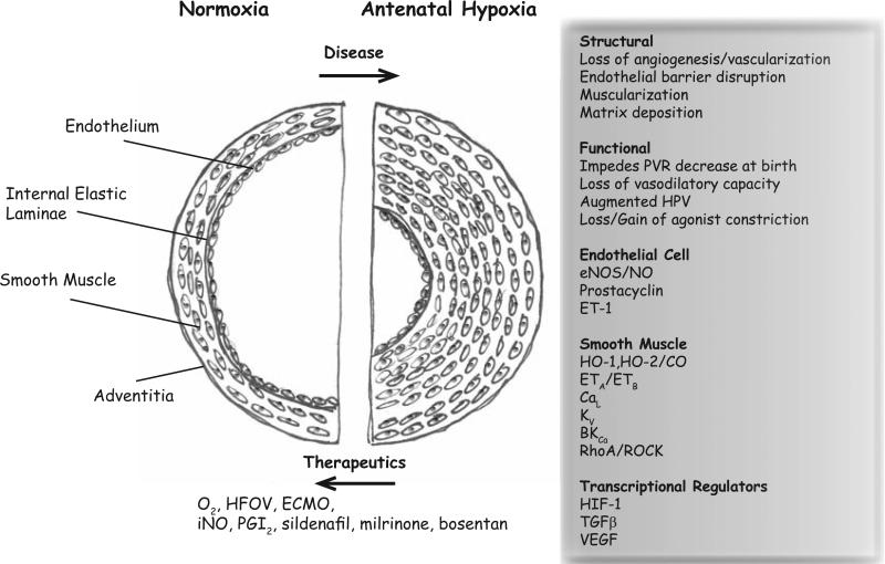
Fig. (1). The pulmonary arterial wall in normal and antenatal hypoxia diseased lung.
In a normal lung the vessel wall and smooth muscle layer is thin. The endothelium lines the lumen of the artery and in distal arteries there is no smooth muscle or elastic lamina. With antenatal hypoxia there can be thickening of the smooth muscle layer that impinges on the arterial lumen along with alterations in myocyte reactivity, as well as disruption of endothelial cell structure with loss of barrier function. These changes are manifested through a number of disruptions involving transcriptional regulators, signaling pathways, and ion channels. The figure summarizes the major components that are discussed in this review.

