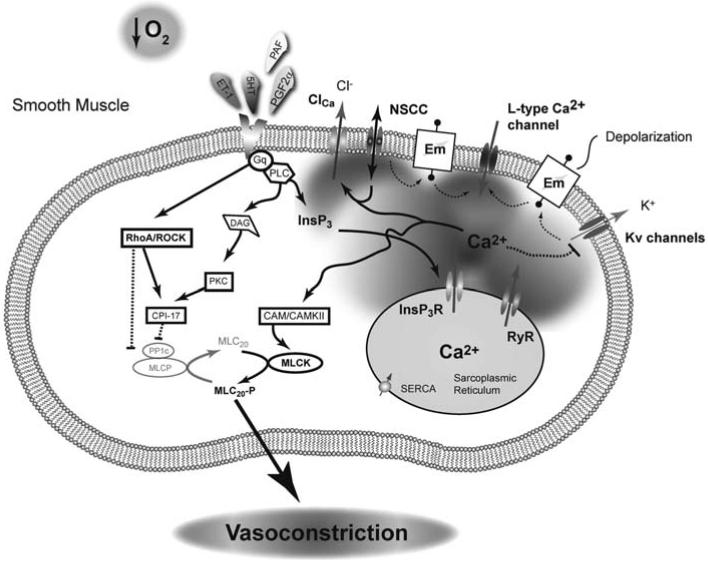
Fig. (2). Pulmonary vasoconstriction in the fetus is a highly coordinated process.
A combination of low oxygen tension and humoral mediators constrict the lung in-utero. The mechanisms associated with hypoxic-induced pulmonary vasoconstriction remain controversial, but a combination of activation of L-type Ca2+ channels, ryanodine receptors, rho-kinase, non-selective cation channels, and inhibition of K+ channels are each important in contraction of pulmonary arteries from adult animals. Pulmonary vascular resistance of the fetal lung is thought to be maintained at a high level due to increased vasoactive agonists, including elevated ET-1 levels. The high ET-1, released from the vascular endothelium, works through a Gq coupled receptor to activate a number of intracellular signaling pathways that lead to smooth muscle cell contraction through simultaneous activation of MLCK and inhibition of MLCP. Solid line with arrow: Activation pathway, Dashed line with bar: Inhibition pathway.

