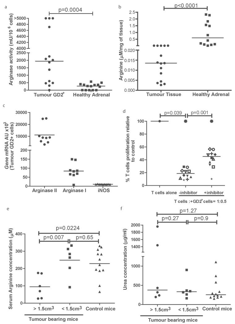
Figure 3. The TH-MYC murine neuroblastoma recreates the arginase dependent microenvironment.
a) Sorted GD2+ cells from murine neuroblastoma tumours have significantly higher arginase activity compared to adrenal tissue. b) Arginine concentrations are significantly lower in the extracellular fluid of neuroblatomas compared to healthy adrenals. c) Expression of Arginase I, Arginase II, and iNOS of GD2+ cells sorted from murine neuroblastoma tumours, by qPCR. d) GD2+ tumour cells from murine neuroblastomas suppress T cell proliferation, with is resorted by arginase inhibition. e) Plasma from tumour bearing mice with large (>1.5cm3) and small (<15.cm3) tumours were analysed for arginine concentration. Plasma arginine is significantly lower in tumour bearing mice with large tumours. f) No increase in plasma arginase activity of tumour bearing mice compared to healthy mice.

