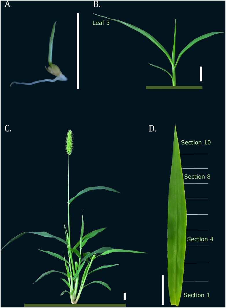
Fig 1. Illustrative figure of each S. viridis tissue and developmental stage that was sampled.
A. Seedling stage (3–5 DAS); B. Young S. viridis, with third leaf fully expanded; C. Mature S. viridis; D. The third leaf 0.5 cm sections that comprised the leaf gradient analysis. Bar corresponds to 1.0 cm.

