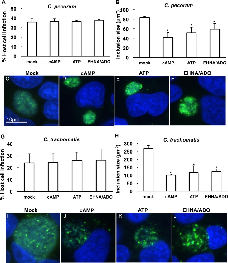
Fig 10. cAMP, ATP and EHNA/ADO inhibit inclusion development less dramatically when added at 14 hpi.
HeLa cells were infected with C. pecorum or C. trachomatis serovar E and exposed to 1 mM cAMP, 1 mM ATP, or 50 μM ADO plus 25 μM EHNA in incubation medium 14 hours after infection. Cells were fixed and labeled with anti-LPS (green) and DAPI (blue) at 35 hours post infection (C. pecorum) or 39 hours post infection (C. trachomatis). Number of inclusions per nucleus was determined and percent infection was calculated for C. pecorum (A) and C. trachomatis (G) (mean ± SD; p > 0.05 in all cases, t test; n = 3). Mean inclusion size was determined for C. pecorum (B) and C. trachomatis (H) (mean ± SD; *p ≤ 0.05, t test; n = 3). Representative images are shown for C. pecorum-infected mock- (C), cAMP- (D), ATP- (E), and EHNA/ADO-exposed (F) cells and for C. trachomatis-infected mock- (I), cAMP- (J), ATP- (K), and EHNA/ADO-exposed (L) cells. Immunofluorescence microscopy images represent one experiment of three independently repeated experiments with similar results.

