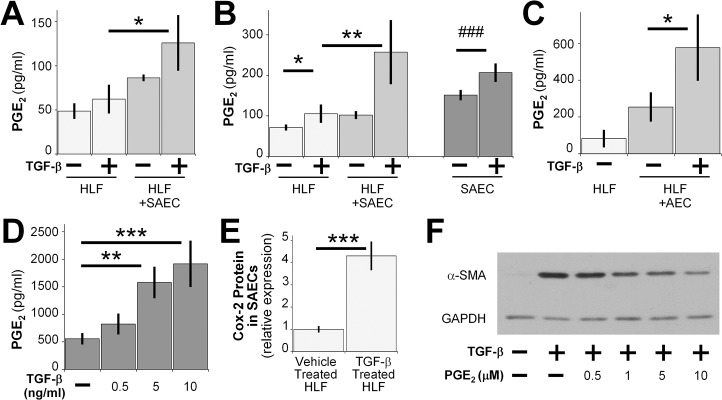
Fig 6. SAECs exert anti-fibrotic effects through the soluble mediator PGE2.
PGE2 in culture medium from SAEC-HLF co-cultures was measured by competitive EIA at 24 hours (A) and 72 hours (B) after TGF-β treatment. (C) PGE2 concentrations in culture medium from AEC-HLF co-cultures were measured at 72 hours. (D) PGE2 in culture medium from SAECs that were treated with TGF-β was measured at 48 hours post treatment. Data shown are mean ± SEM for n = 3 replicates. *** = p<0.001, ** = p<0.01 and * = p<0.05 by ANOVA. ### = p<0.001 by student’s t-test. (E) COX-2 protein expression was analyzed by western blot in SAECs that were co-cultured with HLFs and treated with or without 5ng/ml TGF-β. Densitometry of n = 3 replicates, normalized to untreated control. Data shown are mean ± SD. *** = p<0.001 by student’s t-test. (F) HLFs were treated with or without 5ng/ml TGF-β, or with 5ng/ml TGF-β and increasing concentrations of exogenous PGE2 for 24 hours in serum-free MEM. α-SMA protein expression was analyzed by western blot. A representative blot is shown.

