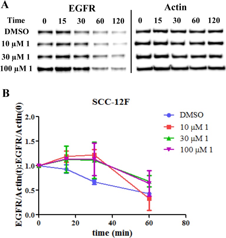
Fig 7. Compound 1 inhibited EGFR degradation in SCC-12F cells.

A. Immunoblots of EGFR at different time points after treatment with 1 at different concentrations or DMSO. Actin served as a loading control. Each experiment included duplicate samples and was repeated in triplicate. B. Time course of EGFR degradation. The ratio of EGFR to actin was quantified at different time points and compared to time zero when ligand EGF was added.
