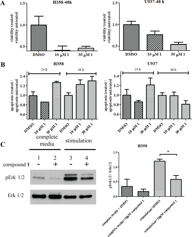
Fig 10. Effects of compound 1 on the Ras-dependent H358 cells.
The U937 FPRΔST cell line was used as control since it had no known Ras-dependency. (A) Compound 1 decreased the viability of H358 cells at 48 h, compared to the control. (B) Apoptosis analysis showed increase response of H358 cells over time compared to control cells. (C) Treatment of compound 1 decreased the phosphorylation level of ERK 1/2 in H358 cells. (-) 0.1% DMSO treated controls; (+) 10 μM compound 1 treated samples. Cells were either grown in complete medium (lane 1 and 2) or stimulated after starvation (lane 3 and 4). The experiment was conducted three times. A representative immunoblot is shown. Immunoblots from all three experiments were quantified by densitometry using exposures in the linear range. The relative ratios of phospho Erk1/2 to total Erk1/2 are plotted. p = 0.0142, calculated with an unpaired two-tailed t-test using GraphPad Prism.

