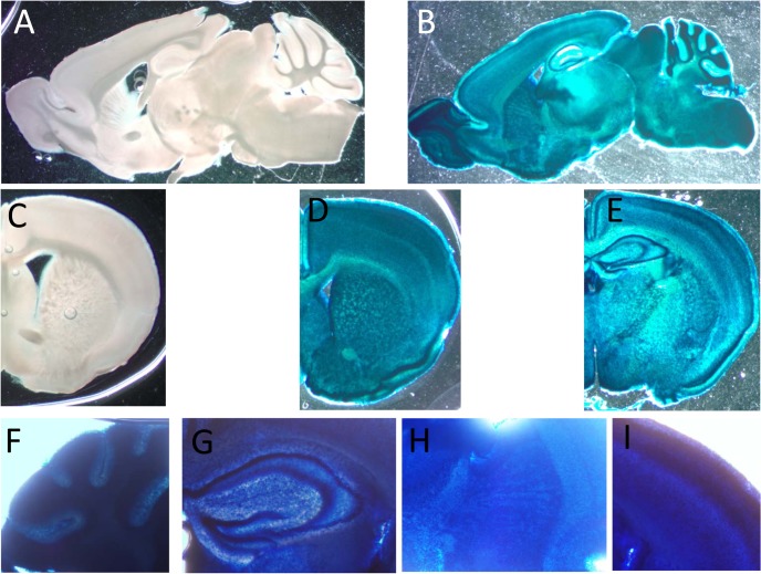
Fig 3. Expression of CaM KMT in the brain of adult C57Bl mice.
Representative staining for β-Gal activity sagittal (B) and coronal (D and E) sections of CaM KMT-/- mice brains, compared to CaM KMT+/+ (A and C). The figure represents results of 4 different adult male C57Bl6/J mice brains. Magnified images of cerebellum lobules (F), hippocampus (G), striatum (H) and M1 region of the cerebral cortex (I) Images exemplify the expression in all cells in all the brain regions. The nuclear staining is attributed to the nuclear localization signal in the β galoctosidase gene.

