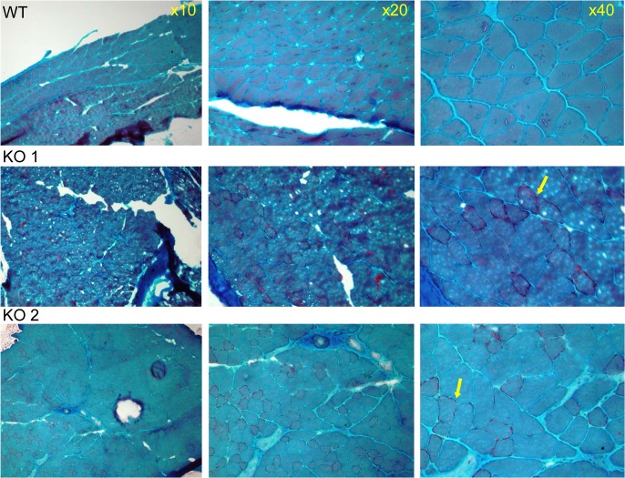
Fig 4. Muscle pathology of CaM KMT-/- mice demonstrated by Gomori-Trichrome staining.
The myopathic feature is shown by the variation in fiber size of the two CaM KMT-/- samples: KO (middle and lower panels), not observed in the sample of the comparable CaM KMT+/+ WT (upper panel). The yellow arrows point the ragged red fibers in the X40 magnification.

