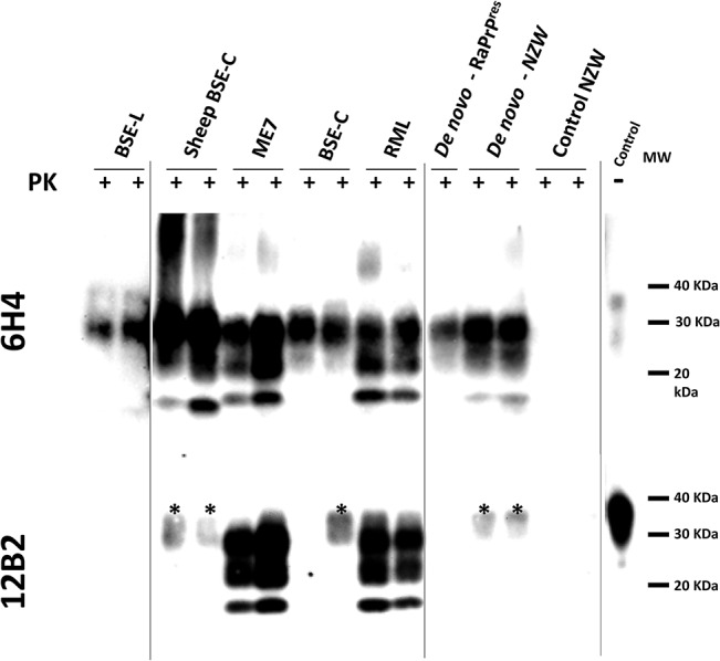
Fig 2. Biochemical analysis of brain homogenates from TgRab mice inoculated with different prion strains.

Two representative brain homogenates (per group) from TgRab mice inoculated with different prion strains [cattle: BSE-L and BSE-C; mouse: RML and ME7; sheep: SSPB/1, sheep BSE-C and atypical scrapie; deer: CWD; rabbit: de novo–RaPrPres (in vitro sample) and de novo–NZW (in vivo sample)] were digested with 100 μg/ml of proteinase K (PK) and analyzed by western blot using two different monoclonal antibodies (upper blot- 6H4 and lower blot– 12B2). Differential electrophoretic migrations and glycosylation patterns observed are consistent with the origin of the prion strains used for inoculation. Control NZW: Normal rabbit brain homogenate. MW: Molecular weight. Vertical lines separate blots with different exposition times.
