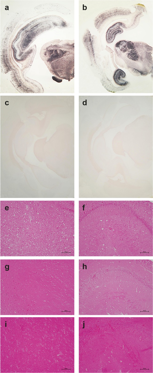
Fig 5. Pathological PrP deposition and vacuolation in the brains of wild-type and tgOv rabbits inoculated with LA21K fast scrapie prions.

Midbrain sections from tgOv (a-b, e-f) and wild-type (c, g-i) rabbits challenged with LA21K fast scrapie prions and from a mock-infected tgOv rabbit (d, i-j). (a-d) PET blot analyses using monoclonal antibody Sha31 showed PrPres accumulation solely in LA21K fast challenged tgOv rabbits. (e-j) Hematoxylin and eosin-stained section at the level of the thalamus (e, g, i) and hippocampus (f, h, j) showing vacuolation, predominantly in the thalamus (e) and to a lesser extend in the hippocampus (f) of the LA21K fast challenged tgOv rabbits, but none in the control animals (g-j). Scale bar: 100 μm.
