
Figure 2.
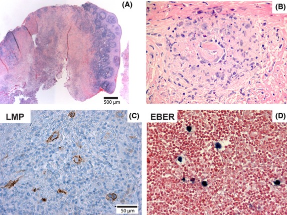
Microscopic findings in an incisional biopsy of EBV-positive oral ulcer. (A) Squamous mucosa with dense lymphoid infiltrate partly covering a large area of necrosis. (B) Small area of viable tissue within the necrotic tissue showing a polymorphous infiltrate that included atypical lymphoid cells. (C) Immunohistochemical staining demonstrated that the atypical lymphoid cells were LMP1-positive (shown here) and CD30-positive (not shown). (D) In situ hybridization for EBER showed scattered positive cells throughout the lesion.