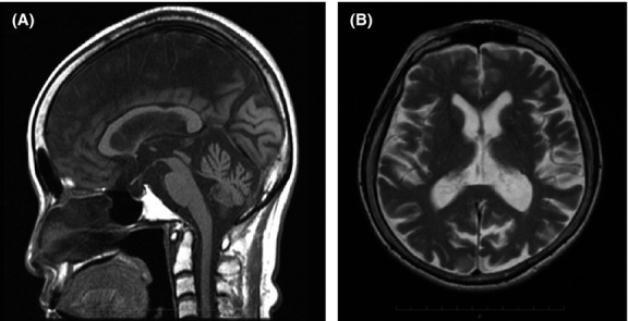
Figure 1.

Brain magnetic resonance imaging findings. (A) T1-weighted sagittal image revealing moderate atrophy of the cerebellar vermis and no atrophy of the midbrain and pons. (B) T2-weighted axial image revealing moderate atrophy of the bilateral cerebellum hemispheres and mild atrophy of the cerebrum. No abnormal high-intensity areas are observed in the cerebrum.
