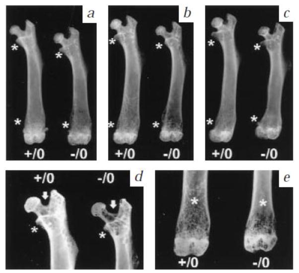
Figure 1.
Radiological analysis of wild type (+/0) and bgn knockout (−/0) bone morphology. Femoral length, bone mass and cortical thickness decreased with age at three (a), six (b) and nine months (c). Detailed high resolution radiographs of 6 months old (d, e) with * indicating regions of reduced trabecular bone mass, and white arrow highlighting the wider angle between the femoral neck and greater trochanter in the mutants. From Xu et al [31] with permission obtained from Nature Genetics.

