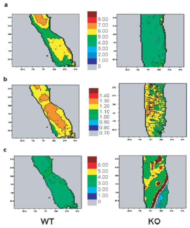
Figure 2.
FTIR imaging of 11-week-old wild type (WT) and SPARC-deficient (KO) mouse femora. a) mineral: organic ratio and b) mineral crystallinity (1030/1020 intensity ratios) are lower in KO while c) collagen maturity is higher compared to WT. From Boskey et al [69] with permission obtained from Journal of Bone and Mineral Research.

