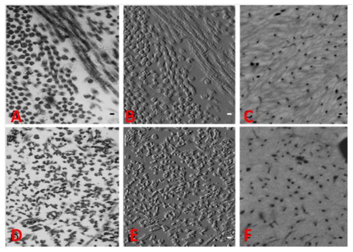
Figure 3.
Electron Microscopy images of 2-month-old wild type (A–C) and Bgn-Dcn−/−;−/− (D–F) mouse tibiae. (B) and (F) are TEM images of (A) and (D) modified for improved visualization of collagen fibril profiles. In Bgn-Dcn−/−;−/− mice the collagen fibrils appear serrated and lack circular cross-sectional profile. The typical collagenous texture seen in WT (C) from quantitative backscattered electron imaging was completely loss in the mutant (F) and replaced by a uniform glassy mineralized matrix. From Corsi et al [43] with permission obtained from Journal of Bone and Mineral Research.

