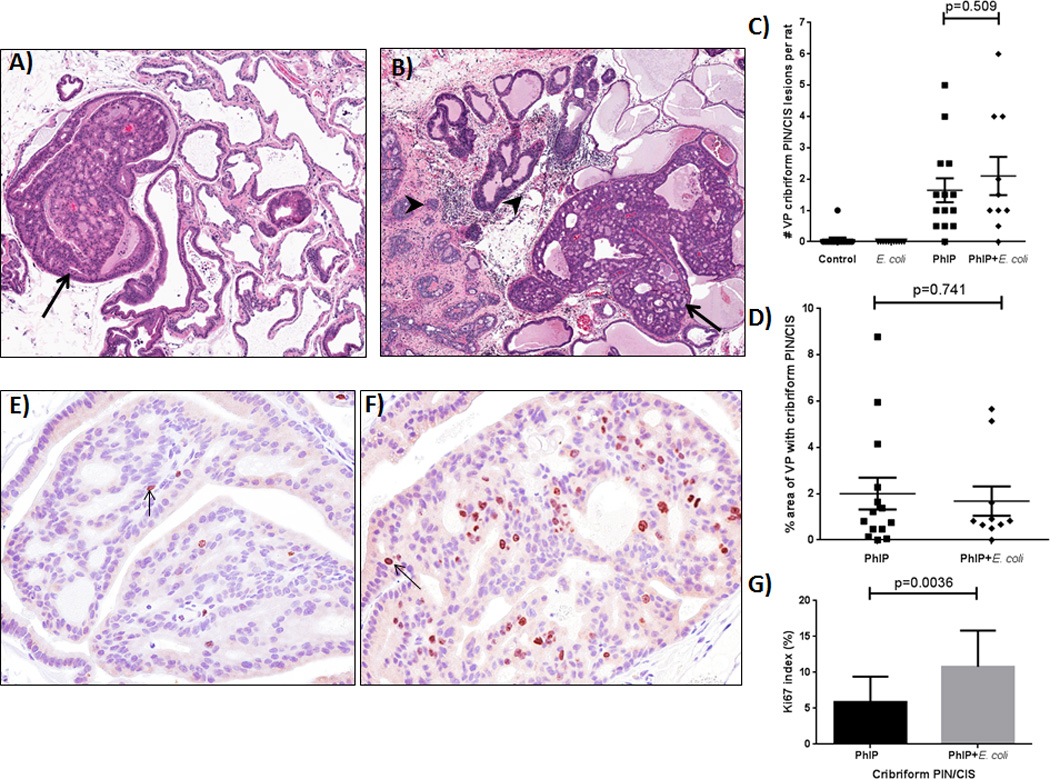
Figure 2.

Development of prostatic cribriform PIN/CIS lesions in PhIP treated animals. Examples of cribriform PIN/CIS lesions (arrows) in the ventral prostate (VP) lobe of PhIP (A) and PhIP+E. coli (B) treated animals. Note chronic inflammation (arrowheads) in PhIP+E. coli treated animal. (C) Number of VP cribriform PIN/CIS lesions observed in study animals. (D) Size of cribriform PIN/CIS lesions as percent area of VP. Two H&E step sections were analyzed per animal and average values are reported. Representative images of Ki-67 IHC in the VP of PhIP (E) and PhIP+E. coli (F) treated animals and Ki-67 index (G).
