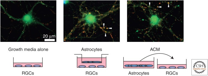
Figure 2.
Retinal ganglion cells (RGCs) can be purified by sequential immunopanning to >99.5% purity from P7 Sprague–Dawley rats and cultured in a neurobasal medium-based growth media that contains several neurotrophic factors, such as brain-derived neurotrophic factor (BDNF) and ciliary neurotrophic factor (CNTF). RCGs are cultured for 3–4 d to allow robust process outgrowth and then cultured for 6 additional d with growth media or astrocyte feeder inserts or with astrocyte-conditioned media (ACM). Change in synapse number in response to treatments is assayed by staining these neurons with antibodies against a pre- and a postsynaptic protein (bassoon, red; homer-1, green). Pre- and postsynaptic proteins appear colocalized (arrowheads) at the synapse because of their close proximity. Astrocytes and ACM strongly increase the number of colocalized synaptic puncta. (Images are courtesy of Sehwon Koh, Eroglu Laboratory, Duke University Medical Center.)

