Figure 2.
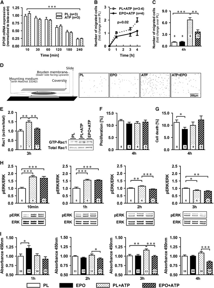
Erythropoietin (EPO) effects on pure microglia cultures. (A) EPO receptor (EPOR) mRNA levels decrease over time, independent of ATP. (B) Increasing number of ATP stimulated microglia (expressed in fold change of the placebo condition) passing through Boyden chamber membranes after 1, 3, and 4 hours, with EPO-treated cells showing less migration (area under the curve P=0.02), (C) most obvious at 4 hours. (D) Boyden membrane slide mount with representative fields-of-view of Boyden chamber membranes of the Hoechst-33342-stained nuclei (inverted) at 4 hours of placebo (PL) or EPO-pretreated microglia ±ATP exposure. (E) EPO pretreatment reduces ATP-stimulated Rac1 GTPase activation at 3 hours; representative western blots. (F) Proliferation or (G) cell death in these cultures. (H) MAPK signaling, investigated by monitoring pERK/ERK after 10 minutes, 1 hour, 2 hours, and 3 hours of 300 μmol/L ATP and 3 IU/mL EPO or PL, shows accelerated inactivation by EPO. (I) EPO decreased microglial water soluble tetrazolium-1 (WST-1) conversion starting with the 2-hour ATP stimulus. Numbers of experiments are given in the bars. Mean±s.e.m. presented, *P⩽0.05, **P⩽0.01, ***P⩽0.001 (two-tailed t-test).

