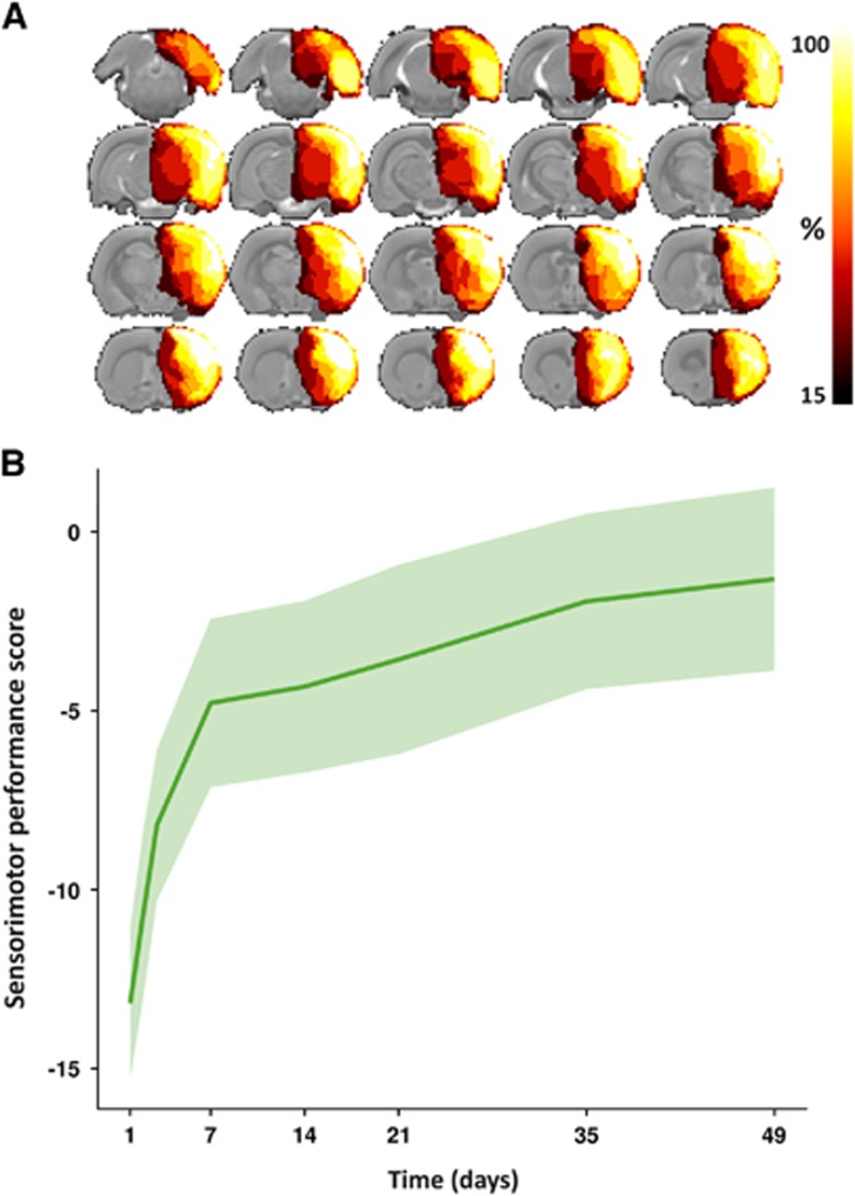
Figure 1.
Removed brain tissue incidence map and sensorimotor performance. (A) Map of the removed tissue incidence after right-sided anatomical hemispherectomy in eight rats, as determined on T2-weighted structural scans. The lesion incidence maps are overlaid on consecutive coronal slices from a multi-slice T2-weighted anatomical rat brain template. The voxel intensities represent the percentage of animals that displayed tissue loss at that specific location at 7 days post surgery. In all animals, ipsilateral cortical tissue was successfully removed. Furthermore, additional removal of (parts of) the CPu and hippocampus was attained in the majority of animals. (B) Sensorimotor performance score as a function of time after hemispherectomy (median (line)±interquartile range (shaded area)). CPu, caudate-putamen complex.

