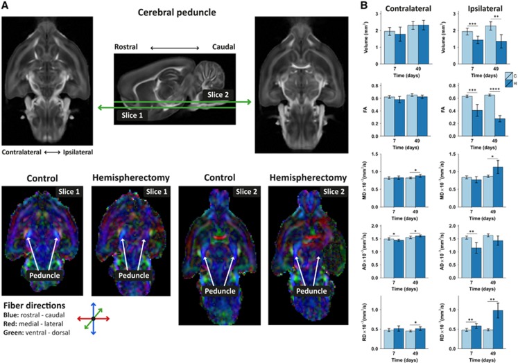
Figure 2.
Ipsi and contralateral cerebral peduncle integrity after hemispherectomy. (A) Examples of diffusion tensor imaging-derived fractional anisotropy (FA) maps of rat brains at 49 days after hemispherectomy and of age-matched controls. Two transversal slices, including the cerebral peduncle, are shown at different ventral–dorsal levels as depicted on the sagittal FA rat brain template image (middle). The cerebral peduncle, running predominantly in rostral–caudal direction displayed symmetrical fiber orientation and FA values in control rat brain (C) ipsi and contralaterally (bottom; slice 1 and slice 2). The FA in the contralateral cerebral peduncle of hemispherectomized rat brain (H) was similar to that in control rat brains, but ipsilaterally it displayed irregular fiber orientation and low FA. (B) Cerebral peduncle regions-of-interest-based volume, FA, mean diffusivity (MD), axial diffusivity (AD) and radial diffusivity (RD), contralaterally and ipsilaterally at days 7 and 49. Bars represent group mean±s.d.; *P<0.05; **P<0.01; ***P<0.001; ****P<0.0001 versus controls as revealed by post-hoc analysis.

