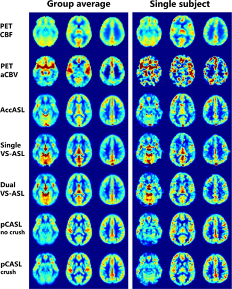
Figure 2.
Example of three transversal slice of the [15O]H2O PET and arterial spin labeling maps of both the group average and a single subject. For comparison of the spatial distribution of the signal, all maps were normalized dividing each voxel by the average gray matter value of the corresponding map. AccASL, acceleration-selective arterial spin labeling; aCBV, arterial cerebral blood volume; CBF, cerebral blood flow; pCASL, pseudo-continuous ASL; PET, positron emission tomography; VS-ASL, velocity-selective ASL.

