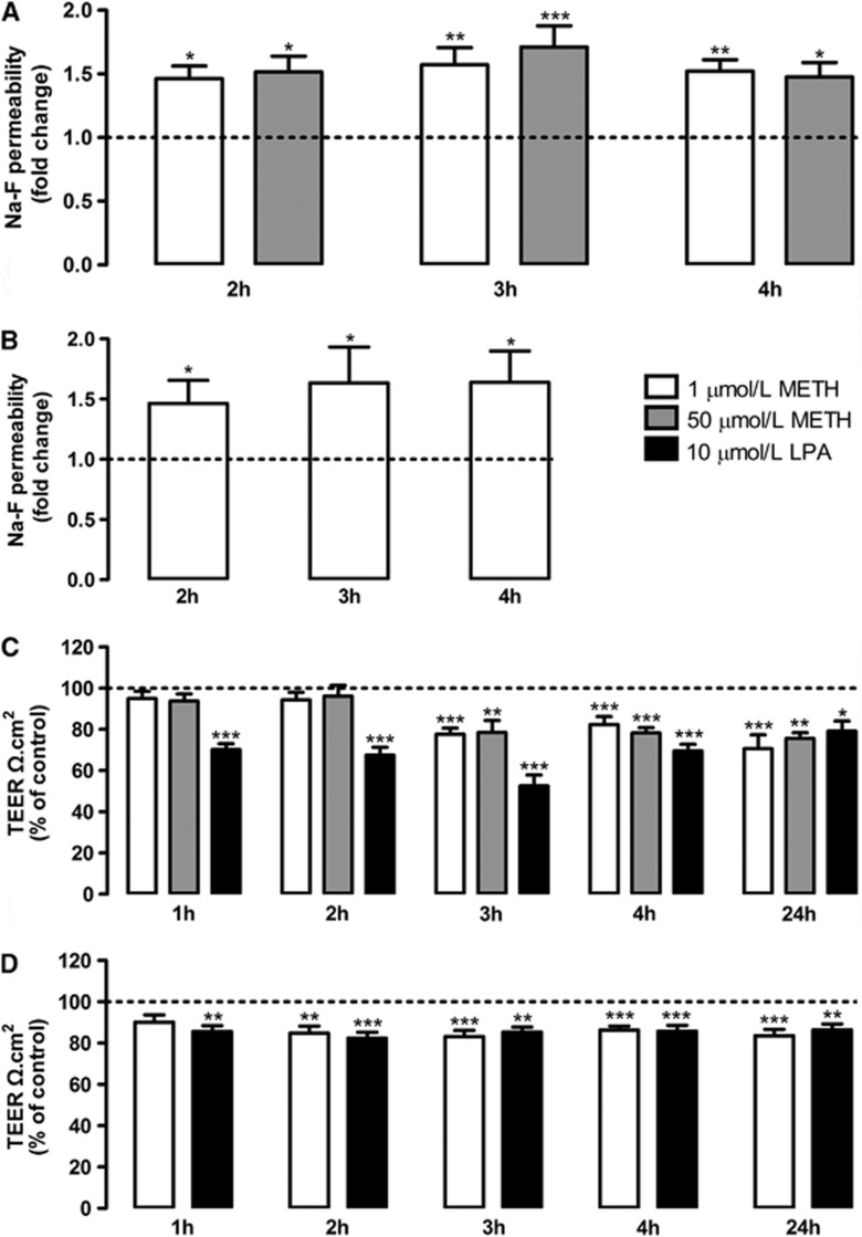
Figure 1.
Methamphetamine (METH) increases the permeability of brain microvascular endothelial cells (BMVECs). (A, B) Macromolecular flux across (A) rat and (B) human BMVECs was assessed using sodium fluorescein (Na-F, 376 Da) at different time points after METH exposure, n=5 to 20. (C, D) Transendothelial electrical resistance (TEER) of confluent (C) rat and (D) human BMVECs monolayers was analyzed under condition of METH or lysophosphatidic acid (LPA) exposure during different time periods, n=5 to 10. All results are shown as means+S.E.M. *P<0.05, **P<0.01, ***P<0.001 significantly different when compared with the control (dashed line) of each time point using Bonferroni's Multiple comparison test.

