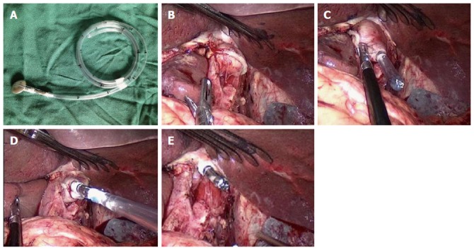
Figure 1.

OrVilTM procedure. A: The central rod of the anvil connected with a tube; B: The lower esophagus was dissociated, and the esophagus was closed and cut; C: The tube of the OrVilTM system was transorally inserted into the esophagus, and the head of the tube was pulled out from the small hole at the end of the esophagus; D: The tube was pulled out until the anvil that connected with the end of the tube reached the esophageal stump; E: The connection line between the tube and anvil was cut down, and the tube was pulled out.
