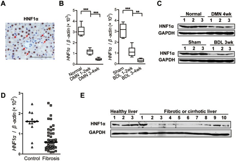
Figure 1.
HNF1α is repressed in fibrotic liver. (A) Immunohistochemical staining of HNF1α in normal rat liver. HNF1α is detected exclusively in the nuclei of hepatocytes (arrow). No obvious staining is observed in non-parenchymal cells (arrow head). Scale bar, 100 μm. (B) mRNA level of HNF1α was assessed by real-time PCR in the livers treated with dimethylnitrosamine (DMN, left) or bile duct ligation (BDL, right) (n = 6 in each group). **P < 0.01; ***P < 0.001 by Mann-Whitney U test. (C) HNF1α protein level in the liver of 3 individual rats after DMN injection (top) or BDL operation (bottom) was detected. (D) A scatter dot plot showing HNF1α expression levels in 12 human control and 44 fibrotic samples as assessed by RT-PCR analysis. Data (median) are normalized to β-actin, and P value was computed by Mann-Whitney U test (P = 0.0008). (E) Western blot analysis of HNF1α in the livers from 3 healthy control individuals and 10 patients with either fibrosis or cirrhosis.

