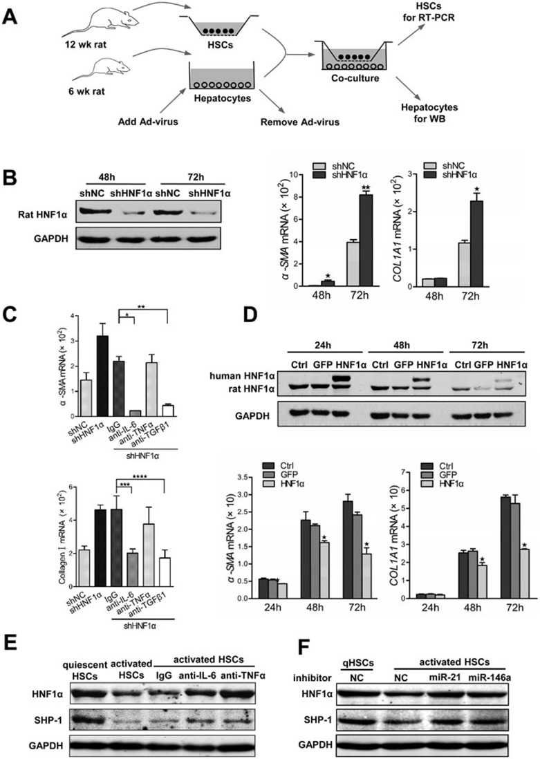
Figure 7.
Crosstalk between HSCs and hepatocytes in vitro. (A) A schematic representation of co-culture experiments with primary HSCs and hepatocytes isolated from rats. (B) Suppression of HNF1α in hepatocytes enhances the activation of HSCs. Endogenous HNF1α level in primary rat hepatocytes pretreated with AdshHNF1α or AdshNC was detected by western blot (left). mRNA levels of α-SMA and COL1A1 in HSCs were assessed by RT-PCR (right). (C) mRNA level of α-SMA and COL1A1 in HSCs co-cultured with AdshHNF1α- or AdshNC-treated hepatocytes. Antibody against IL-6, TNFα or TGFβ1 was added into the co-culture to block the corresponding cytokine. (D) Hepatocytes overexpressing HNF1α attenuates the activation of HSCs. Expression of exogenous human HNF1α and endogenous rat HNF1α in hepatocytes treated with AdHNF1α or AdGFP analyzed by western blot is shown in the top panel; mRNA levels of α-SMA and COL1A1 in HSCs are shown in the bottom panels. (E) Western blot analysis of HNF1α and SHP-1 in hepatocytes co-cultured with quiescent or activated HSCs for 48 h. Antibodies against TNFα, IL-6 and control IgG were used to block the cytokines in co-culture. (F) Expression of HNF1α and SHP-1 in hepatocytes transfected with miRNA inhibitors and then co-cultured with quiescent or activated HSCs for 48 h.

