FIGURE 2.
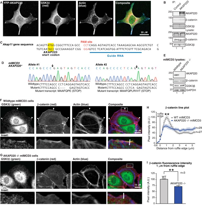
AKAP220 localizes GSK3β and β-catenin to membrane ruffles. A, HEK293 cell expressing YFP-AKAP220 were stained for YFP (AKAP220, green) endogenous GSK3β (red), and actin (phalloidin, blue). AKAP220 and GSK3β co-distribute in the cytosol and are both enriched in membrane ruffles. Scale bar, 20 μm. B, co-immunoprecipitation (IP) of endogenous AKAP220 from wild type mIMCD3 cells detected an interaction with GSK3β and β-catenin. C, CRISPR-Cas9 gene editing was used to disrupt the Akap11 gene using the indicated synthetic guide RNA to the mouse Akap11 gene that encodes AKAP220. D, sequencing analysis of the AKAP220−/− clone demonstrates deletions on both Akap11 alleles (site of deletion indicated by black arrows). The wild type and mutant sequences are listed, with the resulting mutant transcripts from each allele are indicated. Both mutations create early in-frame stop codons. E, complete loss of AKAP220 protein expression was confirmed by Western blot. F, wild type and AKAP220−/− mIMCD3 cells were stained for endogenous GSK3β (green), β-catenin (red), and f-actin (phalloidin, blue). Membrane ruffles were identified as regions with positive f-actin stain. The location of zoomed insets is indicated (white box). In wild type cells, both GSK3β and β-catenin were enriched in ruffles (arrowheads). G, no GSK3β or β-catenin enrichment was observed in membrane ruffles of AKAP220−/− cells. Average peak intensity occured at 1 μm from the ruffle edge in both wild-type and AKAP220−/− cells. This peak intensity was significantly higher in the wild-type cells compared to AKAP220−/− cells (p < 0.01). H, representative quantification of β-catenin distribution at the membrane from one experiment. Fluorescence intensity was measured by line plot analysis, with lines drawn perpendicular to the ruffle edge (representative white lines are indicated on insets). A.U., arbitrary units. I, average β-catenin fluorescence intensity 1 μm from the ruffle edge was significantly reduced in AKAP220−/− cells (p < 0.01; error bars represent S.E.). In wild-type cells, the average intensity 1 μm from the ruffle edge was 101.1 ± 1.9 A.U. In AKAP220−/− cells, the average intensity at the ruffle edge was 71.12 ± 4.1 A.U. (average of n = 3 experiments, >18 cells per condition per experiment).

