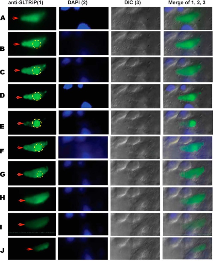
FIGURE 5.
z stacks of a 12-h-old EEF-infected host cell. The infected cell was stained with anti-SLTRiP antibody, and fluorescent images were captured on an Axio-Imager M2 (Zeiss) fitted with ApoTome. The figure shows the export of SLTRiP into the cytosol of the infected host cell (red arrows). Parasite-associated fluorescence is marked with a dotted circle; anything outside the circle is due to exported SLTRiP. In this image, when the parasite is in focus, the exported protein is faintly visible (stack E). Conversely, the exported protein is clearly visible in some stacks, but the parasite is not visible (stacks A and I). A total of 20 z stacks were captured per event, and each stack was 0.2 μm thick. Figure shows only the images from the alternate z stacks. DIC, differential interference contrast.

