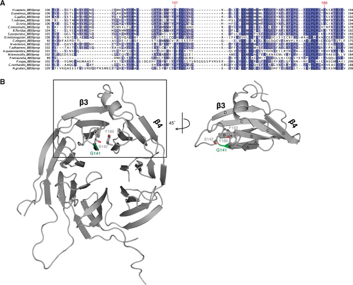
FIGURE 4.
Location of Gly141 in HsBBS91–407. A, fragmentary MSA of BBS91–407 depicting the position and conservation of Gly141, labeled in red. Positions are colored white to blue, according to increasing sequence identity. B, graphic representation of HsBBS91–407 (gray) showing the position of Gly141 (green) and surrounding residues of interest as sticks. The boxed region is rotated by 45° for clarity.

