
FIGURE 2.
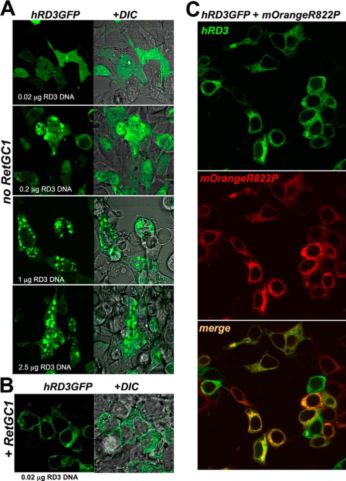
LCA mutation R822P does not block RD3 binding to RetGC1. A, uniform versus punctate pattern of human RD3-GFP localization in HEK293 cells depends on the DNA dose for transfection. RD3 was tagged at the C terminus with GFP as described under “Experimental Procedures,” and RD3-GFP-expressing plasmid was transfected in HEK293 cells using a calcium phosphate precipitation protocol and various amounts of DNA (top to bottom: 0.02, 0.2, 1, and 2.5 μg of DNA cm−2). Left, confocal image of GFP fluorescence; right, the same but superimposed over a differential interference contrast (DIC) image. Note the predominantly uniform distribution of RD3-GFP throughout the cytoplasm and the nucleus at low DNA transfection dose versus the punctate appearance of aggregates in both the cytoplasm and nucleus with the increase in DNA dosage. B, in HEK293 cells expressing low levels of RD3-GFP (0.02 μg of DNA cm−2), its pattern changes to membrane-anchored in the presence of RetGC1 (2 μg of plasmid DNA cm−2 for RetGC1). Note the difference from the pattern in the upper panel of A. C, RD3-GFP co-localizes with the R822P RetGC1. The mixture of 0.02 μg of RD3-GFP plasmid (green) and 2 μg of mOrangeRetGC1 R822P DNA (red) was transfected in HEK293 cells as described under “Experimental Procedures.” Note the difference of the pattern from that of GCAPs in Fig. 1C. The PCC values are presented in Table 1.