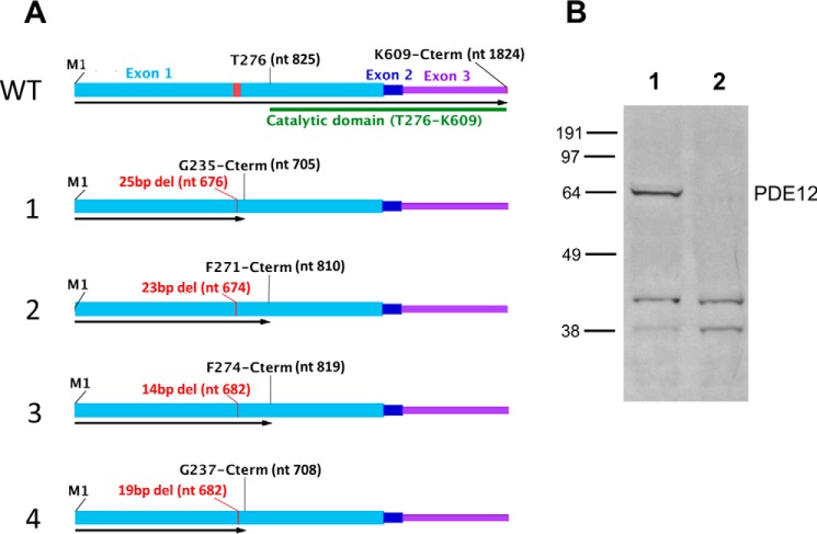
FIGURE 1.
Characterization of frameshift mutations in HeLaΔPDE12 cell line. A, WT, structure of wild-type PDE12 mRNA. Exon 1, light blue; Exon 2, dark blue; Exon 3, purple; coding sequence of EEP nuclease domain, green bar; TALEN nuclease-targeted sequence, red. nt = nucleotide number; M1 = N-terminal methionine; T276 = threonine at the beginning of the catalytic domain. Alleles 1–4, length and positions of the first deleted nucleotide are shown in red; length of open reading frame is shown by a black arrow. Last encoded amino acid residue listed above. B, Western blot detection of PDE12. Lane 1, 40 μg of total cellular protein from HeLa cells. Lane 2, 40 μg of total cellular protein from HeLaΔPDE12 cells.

