FIGURE 8.
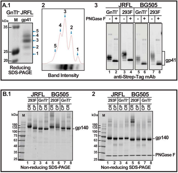
Uncleaved trimers produced in 293F cells are hyperglycosylated. A1, Western blot of reducing SDS gel using the Strep-tag II mAb showing the ladder of gp41 ectodomain bands. A.2, densitometric quantification of the intensity of the gp41 ladder bands shown in A.1. A.3, Western blot of reducing SDS gel using Strep-tag II-specific mAb. B.1, non-reducing SDS gel of uncleaved (CR) and cleaved (CP) JRFL and BG505 gp140 trimers produced in 293F or GnTI− cells. B.2, non-educing SDS gel of samples from B.1 after treatment with PNGase F. The gels were stained with Coomassie Blue. Lanes M, Mr markers. The molecular masses in kDa of marker proteins are shown on the left.

