
FIGURE 2.
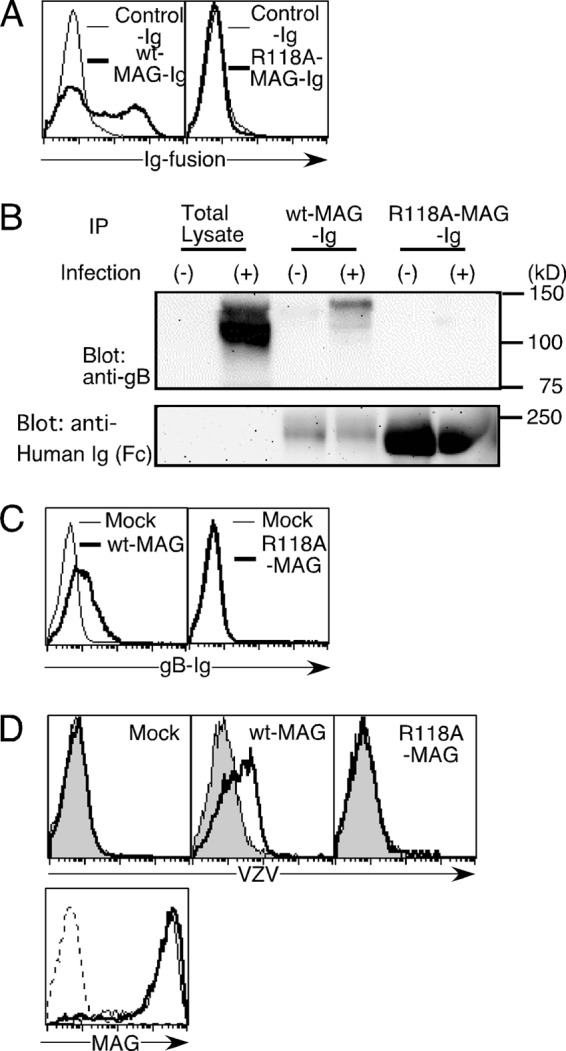
The role of the arginine residue at MAG position 118 in MAG association with gB. A, 293T cells transfected with gB were stained with WT-MAG-Ig (bold line, left panel), R118A-MAG-Ig (bold line, right panel), or control Ig (thin lines, both panels). B, VZV-infected MeWo cells (+) or non-infected MeWo cells (−) were lysed, and the cell lysates were immunoprecipitated (IP) with the indicated Ig fusion proteins. The precipitants and total lysates were analyzed by non-reducing SDS-PAGE, followed by blotting with anti-gB mAb (top panel) or anti-human-Fc Ab (bottom panel). C, 293T cells transfected with WT-MAG (bold line) or mock-transfected (thin line) were stained with gB-Ig (left panel). 293T cells transfected with R118A-MAG (bold line) or mock-transfected (thin line) were stained with gB-Ig (right panel). D, 293T cells transfected with WT-MAG and R118A-MAG and mock-transfected were incubated with VZV virions, followed by staining with anti-gB mAb and secondary antibody. (bold lines). Cells were also stained with antibodies only (gray area, top panels). The relative cell numbers of MAG expression of 293T cells transfected with WT-MAG (thin line) and R118A-MAG (bold line) and mock-transfected (dotted line) are shown (bottom panel).