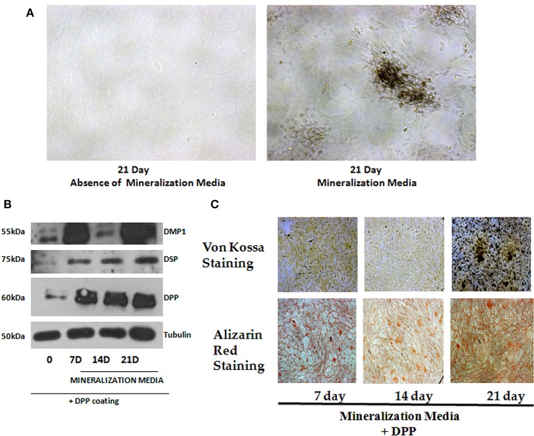
Figure 5.
Role of DPP in terminal differentiation of T4-4 cells. (A) T4-4 cells were cultured on DPP-coated plates in mineralization medium for 7, 14, and 21 days. Light microscopic images of the cells in the presence and absence of mineralization medium at 21 days were taken. Morphological changes in the cells were noticed in the presence of DPP and mineralization medium. (B) Total proteins were isolated from the DPP substrate adherent T4-4 cells and grown under mineralization conditions for 7, 14, and 21 days. Immunoblot analysis was performed for DPP, DSP and DMP1. (C) Alizarin Red and von Kossa staining were performed on T4-4 cells cultured on DPP substrate for 7, 14, and 21 days, respectively.

