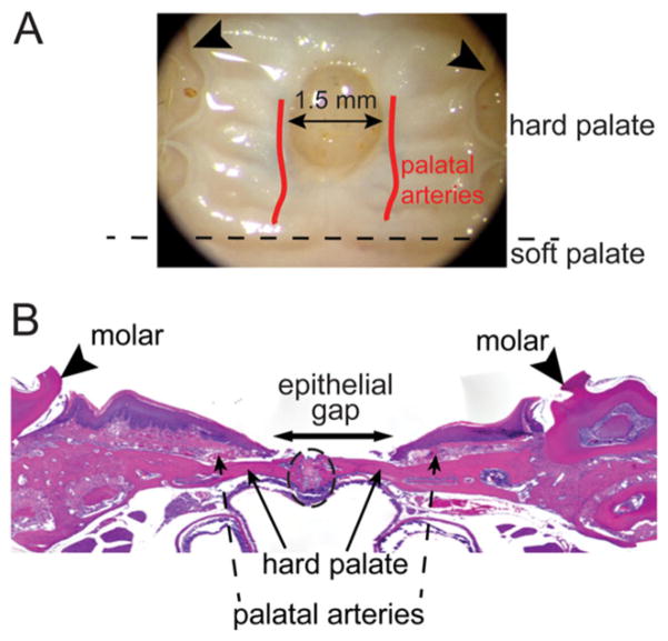
Figure 1.
Schematic of palatal wounding model and quantification of wound healing. (A) Palate wounds were created in the hard palate. The wound site was standardized such that the anterior edge of the wound abutts an imaginary line drawn between the 1st molars (arrowheads) as anatomic reference. Wounds (1.5 mm) are created using punch biopsy dipped in India ink to mark the wound. Both epithelial and submucosal tissues are excised and care is taken to minimize injury to the underlying periosteum and the palatal arteries. (B) H&E stained plate wound sections are used to quantify wound healing phenotype. The inclusion criterion of the wounds in analysis was to make sure that the palate suture line (enclosed within the black dotted oval) is intact with no significant presence of polymorphonuclear leukocytes. Epithelial gap, measured as distance between the encroaching epithelial margins (arrow), is measured to quantify wound closure.

