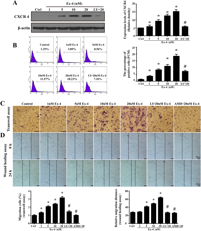
Figure 4. Ex-4 improved CXCR4 expression and migration in MSC via PI3K/Akt.
(A) CXCR4 expression after Ex-4 (0–20 nM) treatment for 24 h was analyzed by western blotting. (B) The flow cytometry was used to quantificational measure the CXCR4 expression. The values on the plots are representative of one experiment. (C) The MSC migration response to SDF-1 was analyzed using a transwell assay. (D) A wound-healing assay was conducted to detect cell motility after MSC were stimulated with or without Ex-4. Bar, 50 μm; *P < 0.05 vs. control group; #P < 0.05 vs. Ex-4 group; LY, LY294002; AMD, AMD3100.

