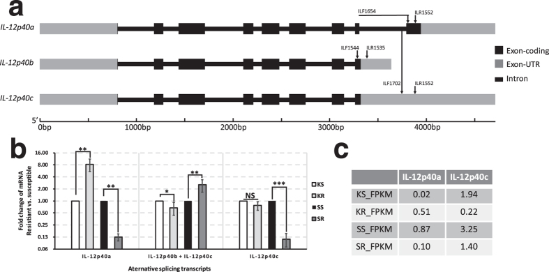
Figure 5. Splicing transcripts of IL-12p40 gene and their expressions in the head-kidney and spleen of C. idella.

(a) Illustration of gene structures of the splice variants. Primers for RT-qPCR validation of the gene expression are showed on the corresponding structures. Primer ILF1654 is designed at the junction of Exon 8 and Exon 9; (b) Fold changes of the splice variants in KR (comparing to KS) and SR (comparing to SS), which are tested by RT-qPCR. *t-test P value < 0.05 ** t-test P value < 0.01; ***t-test P < 0.001; ‘NS’, not significant, t-test P > 0.05. (c) The FPKM values of IL-12p40a and IL-12p40c in the RNA-seq dataset.
